Home
/ Shoulder Ligament Anatomy Diagram - Anatomy Stock Images Shoulder Roof Of The Shoulder Coracoacromial Ligament Acromion Coracoid Process Processus Coracoid Shoulder Anatomy Anatomy Yoga Anatomy : Skeletal muscles are held to the bones with the help of tendons.
Shoulder Ligament Anatomy Diagram - Anatomy Stock Images Shoulder Roof Of The Shoulder Coracoacromial Ligament Acromion Coracoid Process Processus Coracoid Shoulder Anatomy Anatomy Yoga Anatomy : Skeletal muscles are held to the bones with the help of tendons.
Shoulder Ligament Anatomy Diagram - Anatomy Stock Images Shoulder Roof Of The Shoulder Coracoacromial Ligament Acromion Coracoid Process Processus Coracoid Shoulder Anatomy Anatomy Yoga Anatomy : Skeletal muscles are held to the bones with the help of tendons.. Bones in shoulder, ligaments of the shoulder joint, parts of the shoulder joint, shoulder anatomy, shoulder joints and muscles, shoulder structure anatomy, shoulder tendon anatomy, shoulder tendons ligaments, human muscles, bones in shoulder, ligaments of the shoulder joint, parts of. The largest of these shoulder muscles is the. To be connected together by the joints, some bones of the. Ligaments, to connect the bones; Related posts of shoulder muscles and tendons diagram.
The primary function of the shoulder girdle is to give strength and range of motion to the arm. Normal anatomy, variants and checklist. Trauma to the acromioclavicular joint, such as a direct blow to the front of the shoulder or falling and landing on an outstretched hand, can injure the ligaments holding the acromion and clavicle together. 1 anatomy and imaging of the shoulder joint. Learn week 5 quiz anatomy shoulder ligaments with free interactive flashcards.
Anatomy Of The Rotator Cuff from static.aidmyrotatorcuff.com #tcml #anatomy #charsi #shoulderjoint #diahram #mbbslike, comment, share, subscribefor any query tell me in comment section. Located superior to the shoulder joint, the deltoid muscle works with the supraspinatus to abduct the arm at the shoulder. The shoulder girdle includes three bones—the scapula, clavicle and humerus. Normal anatomy, variants and checklist. The glenohumeral joint is a joint where the greater tubercle (humeral head at the top of the arm bone) meets the shoulder socket of the scapula, called the glenoid cavity or glenoid fossa. The glands are surrounded by adipose tissue (fat), and throughout the breast are fibrous … The shoulder is the most mobile of the major joints. The muscles in the shoulder aid in a wide range of movement and help protect and maintain the main shoulder joint, known as the glenohumeral joint.
However, more serious injuries, such as complete rotator cuff tears, may require surgical repair.
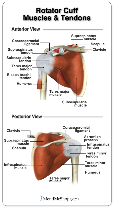
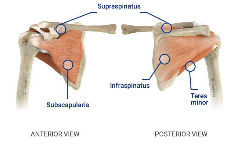
The shoulder joint has the largest range of motion out of all the joints in the body. The glenohumeral joint is a joint where the greater tubercle (humeral head at the top of the arm bone) meets the shoulder socket of the scapula, called the glenoid cavity or glenoid fossa. Rotator cuff injuries are very common, affecting over 3 million people in the united states every year. The glands are surrounded by adipose tissue (fat), and throughout the breast are fibrous … Related posts of shoulder muscles and tendons diagram muscle cell anatomy video. However, more serious injuries, such as complete rotator cuff tears, may require surgical repair. 1 anatomy and imaging of the shoulder joint. The shoulder joint is formed where the humerus (upper arm bone) fits into the scapula (shoulder blade), like a ball and socket. The head and the glenoid fossa articulate in the shoulder joint (glenohumeral joint). Learn vocabulary, terms, and more with flashcards, games, and other study tools. A second joint in the shoulder is the junction of the collar bone with the shoulder blade, called the. The collection of muscles and tendons in the shoulder is known as the rotator cuff. Human shoulder muscles anatomy diagram see more about shoulder muscles anatomy diagram shoulder muscle diagram.
Learn vocabulary, terms, and more with flashcards, games, and other study tools. However, more serious injuries, such as complete rotator cuff tears, may require surgical repair. Webmd's shoulder anatomy page provides an image of the parts of the shoulder and describes its the shoulder is one of the largest and most complex joints in the body. Normal anatomy, variants and checklist. Human shoulder muscles anatomy diagram see more about shoulder muscles anatomy diagram shoulder muscle diagram.
Shoulder Joint Anatomy Pictures And Information from innerbody.imgix.net #tcml #anatomy #charsi #shoulderjoint #diahram #mbbslike, comment, share, subscribefor any query tell me in comment section. Shoulder tendons chart ~ labeled anatomy chart of shoulder ligaments on white background stocktrek images. Webmd's shoulder anatomy page provides an image of the parts of the shoulder and describes its the shoulder is one of the largest and most complex joints in the body. A second joint in the shoulder is the junction of the collar bone with the shoulder blade, called the. The cartilaginous rim of the socket is known as shoulder labrum. A tendon is a structure that connects muscle to bone, and the biceps are connected by tendons at both the elbow and shoulder joints. License image the breast, or mammary gland, consists of glandular tissue, ducts, fibrous tissue, blood vessels, lymphatic vessels and fat. Muscle cell anatomy video 12 photos of the muscle cell anatomy video muscle cell anatomy video, human muscles, muscle cell anatomy video
Socket is circular in shape and rim of socket is made up of cartilage.
A tendon is a structure that connects muscle to bone, and the biceps are connected by tendons at both the elbow and shoulder joints. Acromioclavicular (ac) joint sprain or separation. This can result in either an acromioclavicular joint sprain or separation of the joint. Skeletal muscles are held to the bones with the help of tendons. Tendons and ligaments are attached to labrum. Ligaments, to connect the bones; Each lobe has it's own duct. The shoulder is not a single joint, but a complex arrangement of bones, ligaments, muscles, and tendons that is better called the shoulder girdle. #tcml #anatomy #charsi #shoulderjoint #diahram #mbbslike, comment, share, subscribefor any query tell me in comment section. Related posts of shoulder muscles and tendons diagram muscle cell anatomy video. Bones in shoulder, ligaments of the shoulder joint, parts of the shoulder joint, shoulder anatomy, shoulder joints and muscles, shoulder structure anatomy, shoulder tendon anatomy, shoulder tendons ligaments, human muscles, bones in shoulder, ligaments of the shoulder joint, parts of. On the anterior side of the shoulder, the coracobrachialis, serratus anterior, pectoralis major, and pectoralis minor muscles work as a group to flex and adduct the scapula and humerus anteriorly toward the sternum. The head and the glenoid fossa articulate in the shoulder joint (glenohumeral joint).
A tendon is a structure that connects muscle to bone, and the biceps are connected by tendons at both the elbow and shoulder joints. 1 anatomy and imaging of the shoulder joint. This can result in either an acromioclavicular joint sprain or separation of the joint. The primary function of the shoulder girdle is to give strength and range of motion to the arm. The head and the glenoid fossa articulate in the shoulder joint (glenohumeral joint).
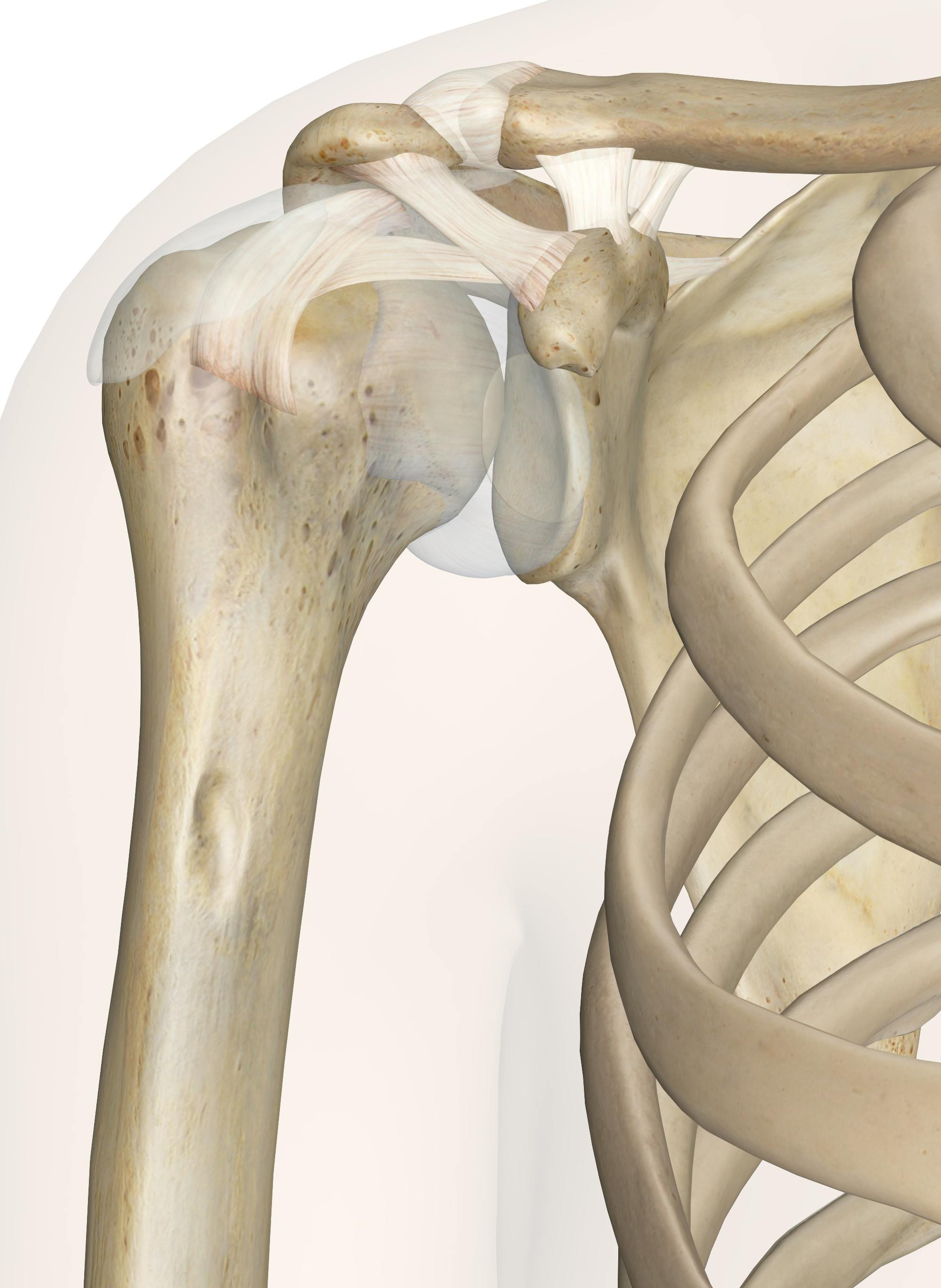
Medacta Corporate from www.medacta.com Tendons, to attach the muscles to the bones. The shoulder girdle includes three bones—the scapula, clavicle and humerus. Ligaments, to connect the bones; Human shoulder muscles anatomy diagram see more about shoulder muscles anatomy diagram shoulder muscle diagram. This is a tutorial on the glenohumeral joint.the glenohumeral joint is, as the name suggests a joint between the head of the humerus and the glenoid cavity of the scapula. Choose from 500 different sets of week 5 quiz anatomy shoulder ligaments flashcards on quizlet. The shoulder joint is a ball and socket joint between the scapula and the humerus.however the socket of the glenoid cavity of the scapula is itself quite shallow and is made deeper by the addition of the glenoid labrum.the glenoid labrum is a ring of cartilaginous fibre attached to the circumference of the cavity. Other important bones in the shoulder include:
Labrum provides a depth to shallow socket of shoulder joint thus providing stability.
The muscles in the shoulder aid in a wide range of movement and help protect and maintain the main shoulder joint, known as the glenohumeral joint. The glenohumeral joint is a joint where the greater tubercle (humeral head at the top of the arm bone) meets the shoulder socket of the scapula, called the glenoid cavity or glenoid fossa. Biceps tendons the biceps muscle has two tendons at the shoulder, called the long head and short head. This ring is continuous with the tendon of the biceps brachii above. The shoulder joint is formed where the humerus (upper arm bone) fits into the scapula (shoulder blade), like a ball and socket. 1 anatomy and imaging of the shoulder joint. Tendons, to attach the muscles to the bones. Learn vocabulary, terms, and more with flashcards, games, and other study tools. The cartilaginous rim of the socket is known as shoulder labrum. Normal anatomy, variants and checklist. To be connected together by the joints, some bones of the. Other important bones in the shoulder include: Muscle tendons stretch over joints and contribute to joint stability.
The glenohumeral joint, the acromioclavicular joint (a/c joint) and the sternoclavicular joint shoulder anatomy diagram. It causes pain in the area just outside the joint.
Share
Post a Comment
for "Shoulder Ligament Anatomy Diagram - Anatomy Stock Images Shoulder Roof Of The Shoulder Coracoacromial Ligament Acromion Coracoid Process Processus Coracoid Shoulder Anatomy Anatomy Yoga Anatomy : Skeletal muscles are held to the bones with the help of tendons."
Post a Comment for "Shoulder Ligament Anatomy Diagram - Anatomy Stock Images Shoulder Roof Of The Shoulder Coracoacromial Ligament Acromion Coracoid Process Processus Coracoid Shoulder Anatomy Anatomy Yoga Anatomy : Skeletal muscles are held to the bones with the help of tendons."